
MRI in the spine is necessary to help make a definative diagnosis and prescribe the proper treatment option. The survey is among the most informative, but requires some preparation and proper interpretation from the results.
INDICATIONS
MRI of the spine is prescribed in almost all cases should there be a suspicion of a pathology from the ridge. The study is desirable for trauma, various developmental abnormalities, inflammatory diseases, degenerative processes, malignant formations, metastases.
The operation is needed:
– in case of severe lumbar pain;
– shooting or aching pains with recoil in the thigh, calf, groin or buttocks;
– incontinence of feces and urine;
– pinching and lack of mobility.
Magnetic resonance imaging is prescribed following the patient may be examined with a neurologist.
WHAT DOES MRI SHOWS?
A radiologist or possibly a doctor of functional diagnostics works with decoding of MRI images of the spine. Three-dimensional cards are weighed against pictures of a proper person, after which possible pathological changes are identified. For instance ,: hernia, osteochondrosis, etc. The analysis might help determine takes place of progression of the condition, as well as pick the right treatment methods. For the cards, you can clearly begin to see the soft tissues and bones – the bones are painted within a dark color, and the spine is in light colors.
What exactly is DISPLAYED Within the IMAGES?
Many people are thinking about what the MRI from the spine shows. The task will demonstrate the next results:
– the degree of possible injury to the spine, as well as the existing pathologies. It is possible to acknowledge them noisy . stages;
– see neoplasms and possible inflammation in soft tissues;
– to look for the nature and extent of the injury;
– to identify a hernia, tomography shows the protrusion in the muscles and longitudinal ligaments.
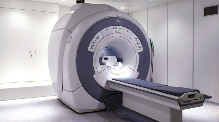
So how exactly does an MRI WORK?
For magnetic resonance imaging, the individual is put inside a special apparatus, the location where the section of ??the body under investigation is scanned using a magnetic field. Facts are saved, printed, visualized, and after that becomes available for analysis by way of a doctor. The method will not cause discomfort, but throughout the MRI you should lie still to the image to get of excellent quality. The research takes about half one hour.
PREPARATION
You’ll want to remove all metal objects: rings, earrings, watches, etc. Cellphones ought to be left outside of the premises. A few hours ahead of the diagnosis, you shouldn’t take food, medications, or drink liquids. It is recommended wear loose-fitting clothing that doesn’t hinder movement. The examination is absolutely painless, and you may eliminate unpleasant sounds in the operation of the tomograph with the help of earplugs.
Contraindications
Absolute contraindications range from the existence of electronic implanted medical devices, ferromagnetic heart valves, the use of massive ferromagnetic medical structures within the body.
Relative contraindications include pregnancy, the use of metal structures inside the skeleton, dentures, prosthetic heart valves, insulin pumps and nerve stimulants.
More info about MRT pozvonochnika you can check the best webpage